CARPAL TUNNEL SYNDROME & OTHER COMPRESSION NEUROPATHIES OF THE UPPER EXTREMITY
- PGH ORTHO
- Nov 6, 2020
- 5 min read
Jose Ma. D. Bautista, MD
Associate Professor in Orthopedics
College of Medicine
University of the Philippines-Manila
Overview
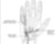
Carpal Tunnel Syndrome (CTS) is a common condition which occurs when the median nerve is compressed at the wrist (Fig. 1). It’s the most common nerve compression in the upper extremity.

Fig. 1. Carpal Tunnel Syndrome (hopkinsmedicine.org)
Etiology
As the carpal tunnel is surrounded by the carpal bones and the transverse carpal ligament, it does not expand. Any narrowing of its dimensions, or increase in content, will lead to an increase in pressure. The increased pressure leads to diminished nerve conduction.
Acute CTS frequently occurs due to a mass effect caused by trauma (wrist fractures/dislocations) and infections (abscesses). Chronic CTS is often idiopathic. Other potential causes would be anatomic (the presence of a mass), systemic (neuropathies, inflammatory conditions), and exertional (repetitive use).
Presentation
Patients with CTS usually complain of numbness and paresthesia involving the volar aspect of the radial 3½ digits of the hand. This is frequently alleviated by shaking/wringing of the hand. As it progresses, there is nocturnal pain and weakness of grip and pinch.
Diagnosis
The use of a hand diagram, where the patient indicates that all or part of the volar surface of the radial 3½ digits is involved is very helpful in differentiating this condition from the other neuropathies which affect the hand (Fig 2). Involvement of the palm should have us consider a compression of the median nerve proximal to the carpal tunnel. Compression of the ulnar nerve will primarily affect the ulnar 1 ½ portion of the hand.

Fig. 2: Katz & Stirrat Hand Diagram. A: Classic findings of Carpal Tunnel Syndrome. B: Probable. C: Unlikely
Diminished sensation and thenar atrophy are the other physical examination findings. Various provocative tests have been described to help us diagnose CTS. These include the Tinel’s sign (Fig. 3), Phalen’s (Fig. 4) and Durkan’s tests(Fig. 5), with varied sensitivity and specificity.

Fig. 3. Tinel’s sign (m.blog.daum.net)

Fig. 4. Phalen’s test (freedpt.wordpress.com)

Fig. 5. Durkan’s test (mitchmedical.us)
Whether electrodiagnostic studies are required to proceed with treatment for patients diagnosed with CTS is still up for discussion. Those who do request for this use it to confirm the diagnosis, rule out additional sites of compression of the median nerve (the “double crush syndrome”), as well as establish the severity and chronicity of the condition.
Xrays are done to rule out any skeletal abnormalities which may cause the condition. Other imaging modalities (Ultrasound and MRI) are not routinely used.
Differentials
The other common compression neuropathy of the upper extremity would be the Cubital Tunnel Syndrome. This involves the ulnar nerve which may become compressed within the sleeve of tissue behind and adjacent to the medial epicondyle of the humerus (Fig. 6). As compared to the Carpal Tunnel Syndrome, there is diminished sensation involving the entire ulnar 1 ½ of the hand (both dorsal and volar) and weakness of the ulnar nerve innervated muscles which move the hand, both intrinsic and extrinsic.

Fig. 6. The Cubital Tunnel (kleinertkutz.com)
A less common condition affecting the ulnar nerve is the Ulnar Tunnel Syndrome, where the nerve is compressed at the level of the wrist within the Guyon’s canal (Fig. 7). This frequently occurs due to a space-occupying lesion which compresses the nerve. Unlike in the Cubital Tunnel Syndrome, when the ulnar nerve is compressed at the wrist, distal to the branching off of the dorsal sensory branch of the Ulnar Nerve, sensory loss only involves the volar aspects of the ulnar 1 ½ fingers and weakness may be observed in the ulnar nerve innervated intrinsic muscles of the hand. The muscles of the forearm innervated by the ulnar nerve are not affected.

Fig. 7. The Ulnar Tunnel (Strohl & Zelouf. JAAOS 2017)
Although involving the same nerve, compression of the median nerve at a different level will result in a presentation different from CTS. Compression of the nerve at the forearm leads to the Pronator Syndrome. This presents with predominantly sensory symptoms. Unlike the CTS, there is sensory loss at the palmar aspect of the thenar eminence. Compression of the Anterior Interosseous Nerve leads to motor symptoms, specifically weakness of the Flexor Pollicis Longus and the Flexor Digitorum Profundus to the index finger. The condition is called the Anterior Interosseous Nerve Syndrome.
The “double crush syndrome” is compression of the median nerve at different levels (such as at the wrist and the cervical root level. The significance of detecting its presence in a patient diagnosed with CTS is that treatment (including surgery) directed towards the CTS will not completely relieve the patient’s symptoms.
Although even less common than the previously mentioned conditions, the radial nerve or its branches may likewise be compressed. As the branches are either predominantly sensory or motor, they will manifest differently. Involvement of the superficial branch of the radial nerve, such as in Wartenberg Syndrome, when the nerve is compressed at the distal forearm, will cause pain and paresthesia at the dorso-radial aspect of the hand (Fig. 8). The Tinel’s sign can be elicited.

Fig. 8. Wartenberg Syndrome (pocayo.com)
The Posterior Interosseous Nerve Syndrome, where this branch of the radial nerve is compressed as it passes through the radial tunnel in the proximal forearm will cause weakness of finger extension, the “finger drop” (weakness of the extensors or inability to extend the metacarpophalangeal joint, but not of the interphalangeal joints which are extended by the intrinsic muscles), with no detectable sensory deficit. Pain may be elicitied during resisted forearm rotation. (Fig. 9)

Fig. 9. The Radial Tunnel (physio-pedia. Com)
Treatment
Most patients with CTS, like those with other compression neuropathies of the upper extremity, are started off with conservative treatment. This usually includes activity modification, immobilization and oral medicines (such as NSAIDs). The wrist splint is the most frequently used form of immobilization for CTS (as well as Ulnar Tunnel Syndrome). In Cubital Tunnel syndrome, it’s the elbow immobilized (usually in extension, with either a rigid brace or even just towels wrapped around the elbow). Patients with Posterior Interosseous Nerve Syndrome may be immobilized in a long arm cast with the elbow flexed at 90o, the forearm supinated and the wrist in neutral.
Corticosteroid injections have been shown to provide good relief for patients with CTS. There however is the risk of recurrence after these injections. Steroid injections are not as successful in most other compression neuropathies of the upper extremity.
Surgery, the Carpal Tunnel Release, wherein the transverse carpal ligament is incised, thus decompressing the median nerve, is the gold standard of treatment (Fig. 10).

Fig. 10. Carpal Tunnel release (Ono, Clapham & Chung. IJGM. 2010)
This may be done open or thru a minimally invasive approach (endoscopic). If adequately done before any permanent damage to the median nerve is present, this leads to excellent results. Surgery to decompress the involved nerve is likewise the last resort for the other compression neuropathies of the upper extremity. In addition to decompression, the ulnar nerve may be transposed (or transferred) anteriorly in certain patients with Cubital Tunnel Syndrome (Fig. 11).

Fig.11. Anterior Transposition of the Ulnar Nerve


